
Retina
We are working with both local and international collaborators to understand how different conditions affect retinal haemodynamics.
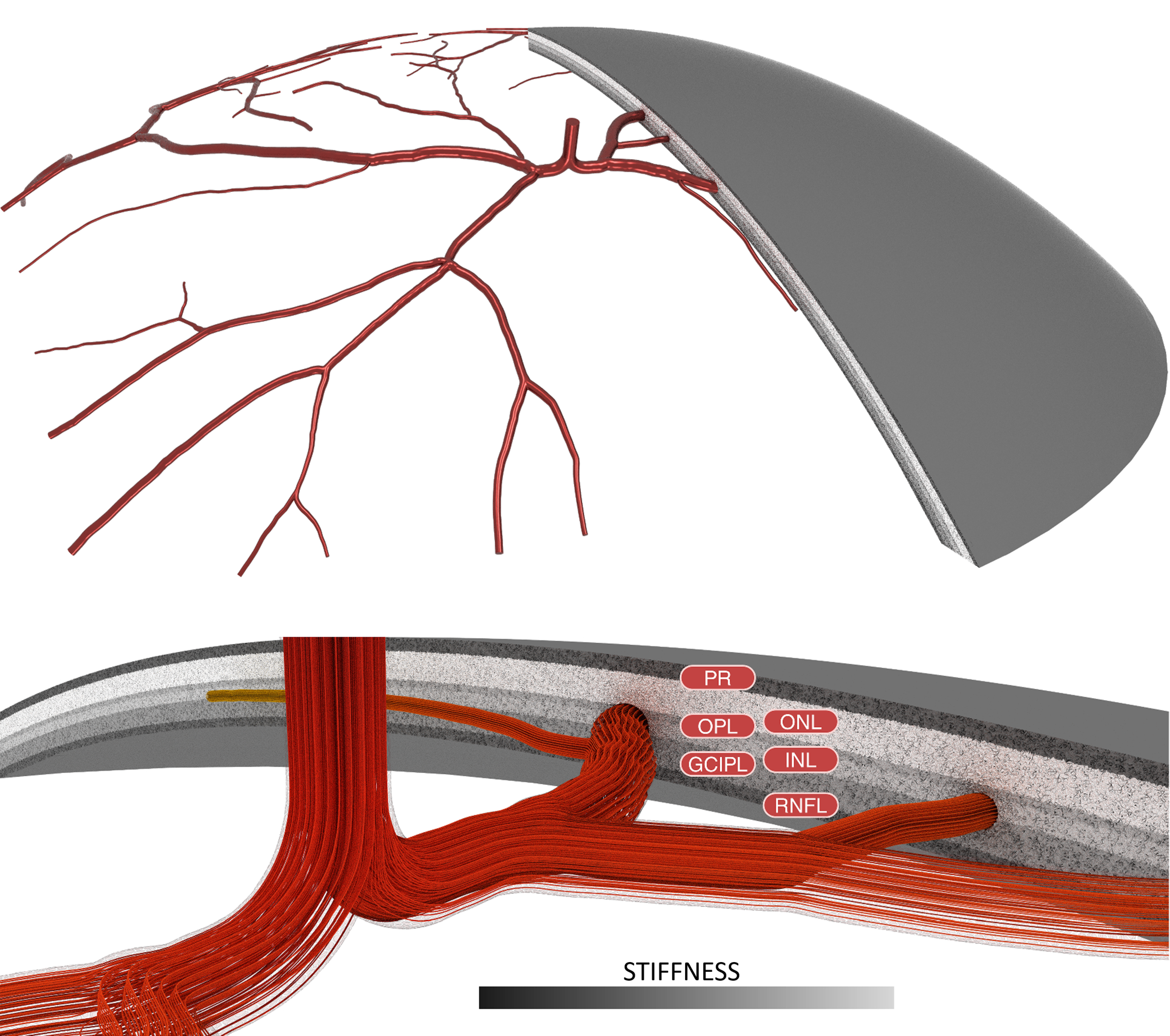
The retina is a layer of light-sensitive tissue found in the posterior segment of the eye. It consists of numerous layers of nerve cells, which a responsible for translating light entering the eye into electrical neural impulses which are transmitted to the brain. As a result, the retina is highly metabolically active, and requires a rich network of microvasculature in order operate effectively.
We use a combination of retinal fundus photography and optical coherence tomography (OCT) to reconstruct subject specific 3D models of the retinal layers and arterial vasculature. We combine this structural information with measured data from Doppler ultrasound in high-fidelity computational fluid dynamics (CFD) simulations.
With this framework we investigate the haemodynamic differences between heathy and diseased retinas (glaucoma and diabetic). We are also investigating conditions of both increased temperature and microgravity to better understand the factors that contribute to a vision degradation condition experienced by astronauts defined as Space Associated Neuro-ocular Syndrome.
